


On March 13, 2025, researchers achieved a groundbreaking milestone in neuroscience and cellular biology by capturing the first-ever image of two PINK1 proteins bound to the surface of a mitochondrion. Using advanced cryo-electron microscopy (cryo-EM), the scientists visualized these proteins in unprecedented detail, marking a major advance in understanding mitochondrial health and its link to Parkinson’s disease.
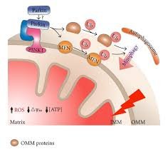
The PINK1 protein (PTEN-induced kinase 1) plays a critical role in mitochondrial quality control. When mitochondria become damaged, PINK1 accumulates on their outer membrane and recruits other proteins, such as Parkin, to initiate the removal of the defective organelle through a process called mitophagy. Mutations in the PINK1 gene are known to cause rare forms of early-onset Parkinson’s disease, a neurodegenerative disorder that affects movement, balance, and coordination.
The high-resolution image captured shows how two PINK1 molecules interact with each other and with the mitochondrial membrane, shedding light on their structural orientation and activation mechanism. This structural insight is expected to fuel drug discovery efforts, helping scientists design targeted therapies that can restore or mimic PINK1 function in patients with Parkinson’s and other mitochondrial disorders.
Experts hail this visualization as a major step forward in mitochondrial biology, made possible by rapid advances in cryo-EM imaging technology, which allows scientists to observe biomolecular structures in their native state at near-atomic resolution.
Marena25
Okay
hossman
No idea about the terms
Suhuyini
Okay
KHANDY
Ok
Walker
Ok
Happy
Good step in technology